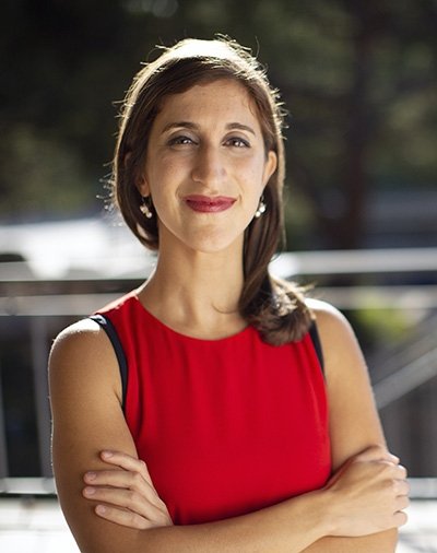
Prof Lisa Poulikakos
Fibrosis, the accumulation of fibrous tissue, is a key feature of many diseases, including neurodegenerative disorders, heart disease and cancer. In oncology, evaluating the extent of fibrosis in a biopsy sample can help determine whether a patient’s cancer is in an early or advanced stage. “The big challenge, however, is that it is extremely difficult to distinguish between these stages using current clinical methods,” said study senior author Lisa Poulikakos, a professor in the Department of Mechanical and Aerospace Engineering at the UC San Diego Jacobs School of Engineering. These methods rely on staining tissues to highlight key structures in the tumor biopsy, but the results can be subjective—one pathologist might interpret a sample differently from another. And while there are more advanced imaging techniques that can provide richer detail, they require expensive, specialized equipment that many clinics simply don’t have.
That’s where the Morpho butterfly comes in. Poulikakos and her team discovered that by placing a biopsy sample on top of a Morpho butterfly wing and viewing it under a standard microscope, they can assess whether a tumor’s structure indicates early- or late-stage cancer—without the need for stains or costly imaging machines. “We can apply this technique using standard optical microscopes that clinics already have,” said Poulikakos. “And it’s more objective and quantitative than what is currently available.”
What are the limitations of current methods for evaluating tumor fibrosis in cancer diagnosis?
Table of Contents
- 1. What are the limitations of current methods for evaluating tumor fibrosis in cancer diagnosis?
- 2. Revolutionizing Cancer Diagnosis: An Interview with Prof. Lisa Poulikakos
- 3. Harnessing Nature’s Beauty for Early Cancer Detection
- 4. An Interview with Prof. Lisa Poulikakos
- 5. Q: Prof. Poulikakos, could you briefly explain the challenge faced in current cancer diagnosis methods, particularly in evaluating tumor fibrosis?
- 6. Q: That’s where the Morpho butterfly comes in. Could you elaborate on how you discovered this unique application?
- 7. Q: How does this method compare to existing techniques in terms of objectivity, quantifiability, and accessibility?
- 8. Q: Looking ahead, what are the next steps for this promising technology?
- 9. Q: Prof. Poulikakos, if you could leave our readers with one thoght-provoking question, what would it be?
- 10. Stay tuned for more groundbreaking research and interviews on Archyde!
Revolutionizing Cancer Diagnosis: An Interview with Prof. Lisa Poulikakos
Harnessing Nature’s Beauty for Early Cancer Detection
 Prof. Lisa Poulikakos, a distinguished figure in the Department of Mechanical and Aerospace Engineering at the UC San Diego Jacobs School of Engineering, has made a groundbreaking discovery that could transform cancer diagnosis. Her innovative use of the Morpho butterfly wing promises a more objective, quantitative, and cost-effective method for assessing tumor stages.
An Interview with Prof. Lisa Poulikakos
Q: Prof. Poulikakos, could you briefly explain the challenge faced in current cancer diagnosis methods, particularly in evaluating tumor fibrosis?
Prof.Poulikakos: “absolutely. Fibrosis is a crucial aspect of cancer progression, and determining its extent in a biopsy sample can definitely help stage the cancer. Though, current clinical methods rely heavily on subjective interpretations of stained tissues. while advanced imaging techniques can provide more detail, they’re often inaccessible due to their high cost and specialized equipment requirements.”
Q: That’s where the Morpho butterfly comes in. Could you elaborate on how you discovered this unique application?
Prof. Poulikakos: “Indeed! We were studying the remarkable optical properties of the Morpho butterfly wing, which exhibits structural coloration without using pigments. We realized that these unique properties could be harnessed to enhance the contrast and detail in our biopsy samples when viewed under a standard microscope. It was a eureka moment when we saw the clear distinction between early- and late-stage cancer structures without the need for stains or expensive equipment.”
Q: How does this method compare to existing techniques in terms of objectivity, quantifiability, and accessibility?
Prof.Poulikakos: “Our method is more objective and quantitative, as it relies on measurable optical properties rather than subjective interpretations of stained tissues. Moreover, it’s highly accessible, as it can be applied using standard optical microscopes that are already present in most clinics. This could considerably improve cancer diagnosis,especially in resource-limited settings.”
Q: Looking ahead, what are the next steps for this promising technology?
Prof.Poulikakos: “We’re currently refining the technique and validating it through larger-scale clinical studies. We’re also exploring its potential applications in other diseases characterized by fibrosis, such as neurodegenerative disorders and heart disease. Our ultimate goal is to see this technology translated into routine clinical use, improving patient care and outcomes worldwide.”
Q: Prof. Poulikakos, if you could leave our readers with one thoght-provoking question, what would it be?
Prof. Poulikakos: “How can we, as a global scientific community, continue to harness the power of nature’s ingenuity to drive medical innovation and improve healthcare for all?”


